Detecting eye and brain disease earlier
The eyes may be the window to the soul, but to scientists, they are also the window to the brain. In particular, the retina, a delicate light-sensing neural network with specialized cells at the back of the eyeball, is linked directly to the brain via the optic nerve and is considered by some to be part of the brain itself.
Now, researchers at the University of Illinois at Chicago are developing imaging techniques that will allow them to study minute changes in the retina that indicate the early stages of brain diseases like Alzheimer’s disease and Parkinson’s disease. Their work is funded by a $1.4 million, four-year grant from the National Institutes of Health National Eye Institute.
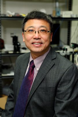
The research, led by Xincheng Yao, the Richard & Loan Hill Professor of Bioengineering and professor of ophthalmology and visual sciences at UIC, will look at how changes in the connections between nerves and blood vessels in the retina — known as neurovascular coupling — are related to the development of eye diseases like
“If we can develop a rapid, noninvasive imaging platform to detect changes in neurovascular coupling that are indicative of eye and brain diseases, we would be able to detect and treat these diseases earlier,” Yao said. “Many times, the only way we can tell there is a disease is once clinical symptoms appear, but the disease process starts long before that. We also have therapies that work best when used early in the disease process, so early detection is an unmet and important need we think our functional optical coherence tomography and OCT angiography (OCTA) technique can help solve.”
Nerves and blood vessels respond to one another, Yao said. When neurons fire or get excited, blood flow increases in the immediate area to bring in glucose and oxygen. Scientists believe that retinal and brain diseases may be presaged by changes in neurovascular coupling that make the vascular response to neural impulses weaker. But detecting these minute changes requires specialized functional imaging systems.
Magnetic Resonance Imaging cannot image individual neurons and blood vessels down to micro capillaries and photoreceptors, according to Yao. In order to image the interactions between nerve cells and capillaries, he will use functional OCT/OCTA, which is able to monitor dynamic interactions of neural and vascular systems, to look at changes in neurovascular coupling in a mouse model of Alzheimer’s disease and a mouse model of retinitis pigmentosa — a rare, progressive disease where the retina degenerates.
“We can’t use functional OCT/OCTA in the brain to look at neurovascular coupling at the cellular level because the brain is surrounded by the skull, and fine resolution isn’t possible,” Yao said. “But we can use functional OCT/OCTA to examine the interactions between individual photoreceptors and their blood vessels in the retina, which is much more accessible in the eye.”
Yao and colleagues will refine their use of functional OCT/OCTA to image neurovascular changes in the retina in the Alzheimer’s mouse model and retinitis pigmentosa mouse model in response to retinal stimulation by light flashes.
Dr. Devrim Toslak and Taeyoon Son from UIC are co-investigators on the grant.
