Researchers use genomics to identify diabetic retinopathy factors
In a search to discover the genetic factors underlying diabetic retinopathy, University of Illinois Chicago researchers also have identified a new approach that can be used as a template to study other diseases.
In the paper, “Integration of genomics and transcriptomics predicts diabetic retinopathy susceptibility genes,” published in eLife, researchers identified genes that respond differently in response to high glucose in individuals with and without diabetic retinopathy.
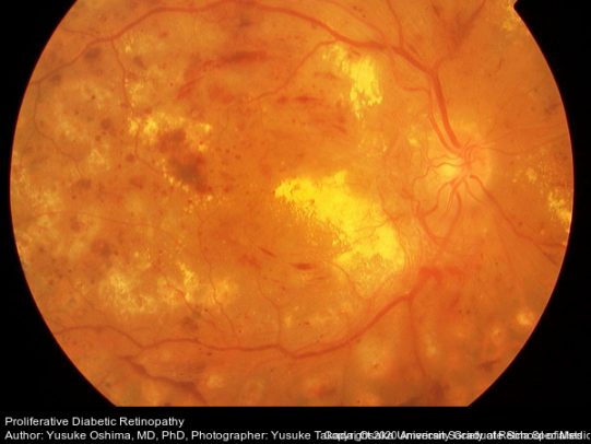
Dr. Michael Grassi, associate professor of ophthalmology at UIC’s College of Medicine, his collaborator, Dr. Barbara Stranger of Northwestern University, and their teams set out to identify genes that cause diabetic retinopathy, a diabetes complication caused by damage to the light-sensitive tissue at the back of the eye — the retina — resulting in vision loss.
Grassi has been interested in diabetic retinopathy since he began his clinical training as a retina specialist.
“I encountered two individuals with disparate outcomes, a 19-year-old who had well-controlled diabetes for five years and went blind, and a Vietnam veteran, who had poorly controlled diabetes for over 30 years but had no vision problems,” Grassi said.
For 10 years, Grassi has been looking at the genetic underpinnings of diabetic retinopathy. After several attempts, he finally landed on a method that resulted in identifying genes that increase the risk of developing retinopathy.
Grassi and his team combined several different methods to identify the gene, known as folliculin, or FLCN, that increases the risk of developing retinopathy. They began by comparing levels of gene activity in individuals with and without retinopathy. A set of genes that was unique to those with retinopathy was identified. Next, they took the genetic markers for this set of genes and found that many were associated with the development of diabetic retinopathy. Finally, they tested whether changes in the levels of some of these genes could cause retinopathy and discovered that increased amounts of FLCN increased the retinopathy risk.
The research team examined glucose-induced changes in gene expression in cell lines from people with type 1 diabetes, both with and without retinopathy. The approach provided new insights into the disease. The identification of single nucleotide polymorphisms, or SNPs, associated with such changes — eQtls (expression quantitative trait loci) — was followed by validation in independent cohorts. The FLCN as a mediator of diabetic retinopathy using Mendelian Randomization further solidified the method.
“It has been a challenge to study diabetic retinopathy because it is so heterogeneous. There are so many genetic factors that can contribute,” Grassi said.
For this study, cell lines generated from blood samples were used from the Diabetes Control and Complications Trial, or DCCT, a large clinical study of diabetic retinopathy. Because the DCCT study generated cell lines for every individual, it allowed for detailed characterization of retinopathy severity in each individual.
Understanding the genetic factors behind diabetic retinopathy can potentially lead to developing new treatment and prevention strategies for retinopathy. The current standard of care involves laser surgery to preserve the center part of vision, or injections into the eye every four weeks.
The research team also includes Ana Marija Sokovic, Poulami Borkar, Amy Lin, Maria Sverdlov, Dingcai Cao, all of UIC; Andrew Skol of the Ann and Robert H. Lurie Children’s Hospital, Chicago; Segun Jung of NeoGenomics Laboratories; Siquan Chen and Sarah Fazal of the University of Chicago; Olukayode Sosina of Johns Hopkins University; Anand Swaroop of the National Eye Institute; and the DCCT/EDIC Study Group.
