Defense grant helps UIC urologist bring prostate cancer into focus
A University of Illinois at Chicago cancer researcher has been awarded a four-year, $560,000 grant from the U.S. Department of Defense to evaluate the usefulness of a novel magnetic resonance imaging technique to discriminate between aggressive and slower growing early-stage prostate cancers.
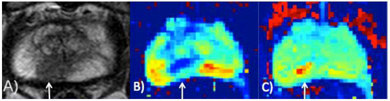
Three images of the same prostate a) Multiparametric MRI b) conventional apparent diffusion coefficient map c) FROC DWI map. The arrow points to a cancerous lesion. Image: Xiaohong Joe Zhou.
Dr. Michael Abern, assistant professor of urology and director of urologic oncology at UIC, will lead a study to evaluate a new MRI technique, developed at UIC by Dr. Xiaohong Joe Zhou, professor of radiology, neurosurgery and bioengineering.
The grant is a Physician Research Training Award designed to help junior faculty clinicians develop strong foundations as physician scientists. Zhou, along with Dr. Peter Gann, professor of pathology at UIC, will serve as co-mentors to Abern for the project. Gann will provide training and assist with tissue analyses to help determine how accurate the new imaging technique is at discriminating aggressive from more indolent cancers. Dr. Andre Balla, director of transdisciplinary pathology and Dr. Vicky Macias, an instructor of pathology in the UIC College of Medicine, will provide expertise in prostate cancer histopathology.
Current methods to diagnose and stage prostate cancer cannot differentiate between aggressive cancers requiring immediate treatment and indolent, or early stage, cancers that can be managed by regular monitoring and periodic re-evaluation, said Abern.
The technique, called fractional order calculus diffusion weighted imaging, or FROC DWI, uses a new approach for detecting the movement of water molecules as they pass through tissues in the body. Typical diffusion MRI is based on the movement of water through the tissue between two points. FROC imaging captures data across multiple points, providing more detail on the architecture of the tissue.
The images show discrete “hot spots” of advanced cancer because water travels through cancer tissue differently than it does through healthy tissue. In tumors, cells are packed more densely and in a disorganized state. As the cancer progresses, the more disorganized and dense its tissue becomes.
“FROC optimizes the ability of MRI to detect differences in the structure and architecture of tissue, which may help us differentiate between indolent and aggressive cancer,” said Abern. The images can be used to guide accurately where to take biopsies, so the likelihood of identifying advanced cancer can be improved.
Standard methods for detecting prostate cancer rely on ultrasound-guided biopsy of the prostate gland. Tissue is removed from 12 different locations throughout the gland and analyzed in the lab. Some facilities use standard MRI to help identify the location of tumors, but these images can’t pinpoint the tumors or give any information on their stage.
“The biopsy procedure might miss cancerous tissue, and the procedure is not very comfortable,” said Abern.
Using standard biopsy, approximately a third of early-stage prostate cancers treated by radical prostatectomy, or removal of the entire gland, turn out to be more advanced. At the same time, 30 percent to 38 percent of men who have a negative biopsy are later diagnosed with prostate cancer in follow-up.
Abern plans to enroll 100 patients with possible prostate cancer based on elevated PSA levels to test whether using FROC DWI to guide biopsies does a better job of detecting aggressive prostate cancer.
All men in the study will undergo standard MRI as well as FROC DWI. They will then receive standard, ultrasound-guided 12-core biopsies, as well as biopsies of targets determined by FROC DWI using fusion technology. Abern will determine which method of guiding biopsies was better at detecting aggressive prostate cancers.
“Some prostate cancers are ‘passengers’, so to speak, that do not put men at significant risk and can be managed through active surveillance,” Abern said. “While others are ‘drivers’ that need aggressive treatment.
“We hope to further test and develop this imaging technique to separate the drivers from the passengers, so we can give men the most appropriate treatment.”
