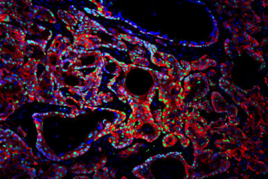
Growing microtumors in a dish helps rapidly identify genes that drive tumor growth
Researchers have identified a new way to screen genes that cause several different types of cancers to grow, identifying particularly promising targets for precision oncology in oral and esophageal squamous cancers.
The study, published in this month’s issue of Cell Reports, used 3-dimensional models of organ tissues called organoids to identify and test potential gene targets from The Cancer Genome Atlas.
“There’s a tremendous amount of data in The Cancer Genome Atlas, and the field has developed life-prolonging and lifesaving precision medicines. But only a minority of these data tell us how cancers grow and whether it’s a drug target,” explained lead author Dr. Ameen Salahudeen, an assistant professor of medicine and biochemistry at the University of Illinois Chicago, and formerly a postdoctoral fellow in the laboratory of Calvin Kuo at Stanford University when the research was initiated. “We needed a scalable, functional method to make heads or tails of the data in terms of what’s driving cancer growth and whether it can be targeted.”
In order to pinpoint the genes that cause tumors to grow, the researchers decided to focus on areas of the genome that displayed two things: regions possessing abnormally high copies of the same gene, something common to many types of cancer, and regions with high levels of RNA expression, which would indicate that those genes are involved in tumor growth. To do this, they used a novel algorithm developed at Stanford by Dr. Jose Seoane, who was at the time in the laboratory of Christina Curtis.
The team identified promising regions within the genome for six different cancers: esophageal, oral cavity, colon, stomach, pancreatic and lung. Next, they built tumor organoids — small tumor tissues in a dish — for each of the six organs and tested their candidate genes on the organoids to see which were associated with growing tumors.
Using organoids for this step is an improvement over previous standard methods, Salahudeen explained. The immortalized cell lines typically used in cellular cancer research often have many additional mutations occurring after years of being grown in the laboratory “that really confound things,” he said. And testing this many potential genes in mice would be unscalable and take years.

In collaboration with Dr. David Root at the Broad Institute and Dr. Bill Hahn at the Dana Farber Cancer Institute, the team then screened the organoids using lentivirus libraries of these genes. The organoids revealed particularly interesting results for squamous cancers of the oral cavity and esophagus, both of which have very few known driver genes that could be targeted with drugs. Furthermore, the researchers tested a clinically available small molecule — a type of drug known as an FGFR inhibitor — on the esophageal organoids and were able to shrink the tumor.
“That gene, FGF3, is amplified in nearly half of esophageal squamous cancers and is known to interact with FGFR,” Salahudeen said. “So this could potentially benefit nearly half of esophageal cancer patients.”
Salahudeen, who is a member of the University of Illinois Cancer Center, said the next step is to investigate existing clinical FGFR inhibitors in this new indication within esophageal squamous cancer patients and to continue investigating other potentially promising genes that the study identified.
Other authors on the paper include co-lead author Kanako Yuki in the Calvin Kuo lab at Stanford. Funding for the study came from the NCI Cancer Target Discovery and Development Network to Kuo and William Hahn; the National Institute for Dental and Craniofacial Research; “LaCaixa” Foundation; Emerson Collective; Ludwig Cancer Research and Stand Up To Cancer to Kuo.
